CAT Scan (CT Scan)
Milford Regional maintains several scanners that take advantage of current procedures and protocols utilizing the lowest radiation dose possible. These scanners provide extremely detailed information about bones, soft tissue and blood vessels.
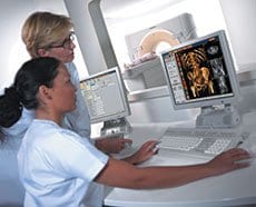
CT (computed tomography), sometimes called CAT scan, uses special x-ray equipment to take pictures of the body from different angles. A computer then processes the information, showing a cross section of body tissues and organs. To learn more about CAT scan, how to prepare for your procedure and what you can expect while having a CAT scan, choose from the list below or speak with your physician.
CAT Scan (CT) – Abdomen
CAT Scan (CT) – Angiography
CAT Scan (CT) – Body
CAT Scan (CT) – Chest
CAT Scan (CT) – Head
CAT Scan (CT) – Sinuses
CAT Scan (CT) – Spine
CAT Scan (Pediatric)
CT Guided Biopsy (Lung)
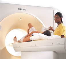
When it comes to heart attack, stroke, trauma or cancer detection, time is of the essence… every second matters. It can mean the difference between life and death or affect the extent of your recovery. When seconds count, you can rely on Milford Regional’s two multi-slice Philips Brilliance Spiral CT scanners.
 A CT scanner takes x-ray images at many different angles through a doughnut shaped, rotating cylinder that patients pass through on a table. Cross sectional images of the body are then produced on a computer. Each image is an X-ray slice of a body structure or organ, which can be analyzed individually or brought together as a three dimensional image. CT scanners provide extremely detailed information about bones, soft tissue and blood vessels.
They are utilized largely to:
- Detect internal injuries and internal bleeding from trauma
- Detect and monitor diseases such as cancer, heart disease, stroke and neurological cases
- Diagnose muscle and bone disorders, such as bone tumors and fractures
- Pinpoint the location of a tumor, infection or blood clot
- Guide procedures such as surgery, biopsy and radiation
The highly detailed images produced by the amazing speed of the multi-slice scanners and increased number of X-ray detectors (taking up to 192 separate images per second) mean radiologists can visualize more, provide earlier detection and begin treatment faster.
Amazingly, scans can now be done in a single breath hold! A full body scan can be done in 30 seconds; the chest and abdomen in 10 seconds and the heart in one-tenth of a second! Scanner speed is significant because any movement can affect image quality. Thus, the scanner must be fast enough to take images of the heart at rest. The exceptional speed also allows radiologist to view blood flow through the arterial and venous vessels of the body as well as the heart.
 Read about how the scanners are used in various modalities.


