Bone Densitometry (Bone Density)
Bone density scanning is an enhanced form of x-ray technology that is used to measure bone loss. It is most often performed on the lower spine, hips and forearm.
This test is most often used to diagnose osteoporosis, a condition that often affects women after menopause but may also be found in men. Osteoporosis involves a gradual loss of calcium, as well as structural changes, causing the bones to become thinner, more fragile and more likely to break.
A bone density scan is effective in tracking the effects of treatment for osteoporosis and other conditions that cause bone loss. It can also assess an individual's risk for developing fractures. The risk of fracture is affected by age, body weight, history of prior fracture, family history of osteoporotic fractures and life style issues such as cigarette smoking and excessive alcohol consumption. These factors are taken into consideration when deciding if a patient needs therapy.
Consult Your Physician
- if you recently had a barium examination or have been injected with a contrast material for a CT scan. If so, you may have to wait 10 to 14 days before undergoing a bone density test.
- if there is any possibility that you are pregnant.
Exam Preparation
On the day of the exam you may eat normally. You should wear loose, comfortable clothing, avoiding belts or buttons made of metal. You will be asked to remove jewelry from the forearm being scanned since it could interfere with the x-ray images.
The Exam
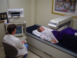
 During the exam, the patient lies on a padded table. An x-ray generator is located below the patient and an imaging device, or detector, is positioned above.
To assess the spine, the patient's legs are supported on a padded box to flatten the pelvis and lower the (lumbar) spine. To assess the hip, the patient's foot is placed in a brace that rotates the hip inward. In both cases, the detector is slowly passed over the area, generating images on a computer monitor.
The machine sends a thin, invisible beam of low-dose x-rays with two distinct energy peaks through the bones being examined. One peak is absorbed mainly by soft tissue and the other by bone. The soft tissue amount can be subtracted from the total and what remains is a patient's bone mineral density.
The bone density test is usually completed within 10 to 20 minutes.
After the Exam
A radiologist will analyze the images and send a report to your primary care physician, or referring physician, who will discuss the results with you.
Your test results will be in the form of two scores:
T score — This number shows the amount of bone you have compared with a young adult of the same gender with peak bone mass. A score above -1 is considered normal. A score between -1 and -2.5 is classified as osteopenia (low bone mass). A score below -2.5 is defined as osteoporosis. The T score is used to estimate your risk of developing a fracture.
Z score — This number reflects the amount of bone you have compared with other people in your age group and of the same size and gender. If this score is unusually high or low, it may indicate a need for further medical tests.
Small changes may normally be observed between scans due to differences in positioning and usually are not significant.

